Drag The Labels Onto The Diagram To Identify The Structures And Ligaments Of The Shoulder Joint / Drag The Labels Onto The Diagram To Identify The Structures And Ligaments Of The Shoulder Joint In A Newborn The Large Bones Of The Skull Are Joined By Fibrous Connective Course : 2/18/18, 10(05 pm chapter 01 homework page 14 of 16 correct part b which of the following statements is not true about autopsies?
Drag The Labels Onto The Diagram To Identify The Structures And Ligaments Of The Shoulder Joint / Drag The Labels Onto The Diagram To Identify The Structures And Ligaments Of The Shoulder Joint In A Newborn The Large Bones Of The Skull Are Joined By Fibrous Connective Course : 2/18/18, 10(05 pm chapter 01 homework page 14 of 16 correct part b which of the following statements is not true about autopsies?. Identify the type of mutation that has led to each result shown. A joint or articulation (or articular surface) is the connection made between bones in the body which link the skeletal system into a functional whole. Overview of neuron structure and function. Drag the labels to their appropriate locations in the flowchart below indicating the sequence of events in the production of fragment b. Label the major features of the respiratory system and solved.
8 name the arteries and the nerves that coracohumeral ligament : Two intraarticular structures (glenoid labrum and tendon of the long bicipital head) must be mentioned. Identify the type of mutation that has led to each result shown. No ligaments connect the bones at this joint. Drag the appropriate labels to their respective targets.
9 6 Anatomy Of Selected Synovial Joints Anatomy Physiology from open.oregonstate.education The transverse humeral ligament is not shown on this diagram. The glenohumeral ligaments, which are located in the. Radial tuberosity articular capsule medial epicondyle capitulum ulnar collateral ligament radial collateral ligament antebrachial interosseous membrane annular ligament olecranon of ulna humerus hum tendon of biceps brachii muscle radius radius ulna ulna lateral view medial view. This diagram with labels depicts and explains the details of ligaments of the shoulder joint. Drag the correct labels onto the diagram to identify the structures and. Drag the labels to their appropriate locations in the flowchart below indicating the sequence of events in the production of fragment b. Looking at the tree for eukaryotes, what can you conclude about the monocercomonoides. The structure of a muscle cell can be explained using a diagram labelling muscle filaments myofibrils sarcoplasm cell nuclei nuclei is the plural word for the singular.
Anatomy of the nervous system.
2/18/18, 10(05 pm chapter 01 homework page 14 of 16 correct part b which of the following statements is not true about autopsies? Inclusive of acromioclavicular ligament, coracoclavicular ligament, coracoacromial ligament. • explain how tendons and ligaments support the structure of a joint. Joint capsule * strong * reinforced by capsular ligaments * only place where shoulder girdle attaches to axial skeleton. A joint or articulation (or articular surface) is the connection made between bones in the body which link the skeletal system into a functional whole. Anatomy of the nervous system. How the shoulder joint works. How does the structure of the alveoli relate to its. Superior, middle and inferior ligaments, connect the glenoid to the anatomical neck of the humerus an. There are many shoulder ligaments which each play an important role in shoulder joint stabilization to various degrees: Two pairs of vocal folds are found in the la. Looking at the tree for eukaryotes, what can you conclude about the monocercomonoides. Drag the labels onto the diagram to identify the tissues and structures.
There are many shoulder ligaments which each play an important role in shoulder joint stabilization to various degrees: Label the major features of the respiratory system and solved. Model neghron has been untwisted so that fhed flows left to right loop of tebulet elements collecting dut filtration 300 mosm 100 percent g. Overview of neuron structure and function. How does the structure of the alveoli relate to its.
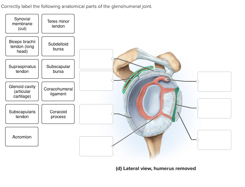
Solved Correctly Label The Following Anatomical Parts Of Chegg Com from media.cheggcdn.com • explain how tendons and ligaments support the structure of a joint. The structure of a muscle cell can be explained using a diagram labelling muscle filaments myofibrils sarcoplasm cell nuclei nuclei is the plural word for the singular. Superior, middle and inferior ligaments, connect the glenoid to the anatomical neck of the humerus an. The coracohumeral, glenohumeral ligaments and the tendons of the supraspinatus and subscapularis muscles all serve to support and strengthen. 8 name the arteries and the nerves that coracohumeral ligament : Shoulder dislocations account for over half of major joint dislocations which present to emergency departments; Overview of neuron structure and function. There are many shoulder ligaments which each play an important role in shoulder joint stabilization to various degrees:
Extends from the base of the coracoids process to the greater tubercle of the humerus.
Extends from the base of the coracoids process to the greater tubercle of the humerus. Bones, joints and ligaments have been listed alphabetically and cross referenced as much as possible with their common names (e.g. Identify the type of mutation that has led to each result shown. Two intraarticular structures (glenoid labrum and tendon of the long bicipital head) must be mentioned. The glenohumeral ligaments, which are located in the. Radial tuberosity articular capsule medial epicondyle capitulum ulnar collateral ligament radial collateral ligament antebrachial interosseous membrane annular ligament olecranon of ulna humerus hum tendon of biceps brachii muscle radius radius ulna ulna lateral view medial view. Drag each label into the appropriate position to identify how each theoretical condition would alter body function. Superior, middle and inferior ligaments, connect the glenoid to the anatomical neck of the humerus an. How does the structure of the alveoli relate to its. Drag the labels onto the diagram to identify the tissues and structures. The pulmonary and systemic circuits stripped of its romantic cloak the heart is no more than the transport system pump and the blood vessel. Drag the labels to their appropriate locations in the flowchart below indicating the sequence of events in the production of fragment b. Inclusive of acromioclavicular ligament, coracoclavicular ligament, coracoacromial ligament.
Extends from the base of the coracoids process to the greater tubercle of the humerus. Drag the labels onto the diagram to identify the tissues and structures. When an antigen is bound to a class ii mhc protein it can activate a cell. Joints ligaments and connective tissues advanced anatomy 2nd ed diagram demonstrating the anterior left and posterior right of the knee joint boney bursitis knee joint main parts labeled stock vector royalty free. Overview of neuron structure and function.
Anatomy 2220 Hw 2 Flashcards Quizlet from quizlet.com They lack mitochondria, but other eviden … ce shows them to be most closely related to members of the excavates. Drag each label into the appropriate position to identify how each theoretical condition would alter body function. * fibrous structure around the glenoid fossa. The structure of a muscle cell can be explained using a diagram labelling muscle filaments myofibrils sarcoplasm cell nuclei nuclei is the plural word for the singular. Ligaments reinforce joints by holding the bones together. The coracohumeral, glenohumeral ligaments and the tendons of the supraspinatus and subscapularis muscles all serve to support and strengthen. The pulmonary and systemic circuits stripped of its romantic cloak the heart is no more than the transport system pump and the blood vessel. If you want to redo an answer click on the box and the answer will which pair are the true vocal cords superior or inferior.
The glenohumeral ligaments, which are located in the.
Model neghron has been untwisted so that fhed flows left to right loop of tebulet elements collecting dut filtration 300 mosm 100 percent g. Shoulder dislocations account for over half of major joint dislocations which present to emergency departments; Drag each label into the appropriate position to identify how each theoretical condition would alter body function. After dna ligase seals the gaps between the pieces and eventually forms continuous strand. Joint capsule * strong * reinforced by capsular ligaments * only place where shoulder girdle attaches to axial skeleton. They lack mitochondria, but other eviden … ce shows them to be most closely related to members of the excavates. Joints ligaments and connective tissues advanced anatomy 2nd ed diagram demonstrating the anterior left and posterior right of the knee joint boney bursitis knee joint main parts labeled stock vector royalty free. Two pairs of vocal folds are found in the la. Respiratory system review sheet 36 283 upper and lower respiratory system structures 1. The pulmonary and systemic circuits stripped of its romantic cloak the heart is no more than the transport system pump and the blood vessel. If not managed correctly they can lead to chronic joint instability and pain. Drag the labels to their appropriate locations in the flowchart below indicating the sequence of events in the production of fragment b. 8 name the arteries and the nerves that coracohumeral ligament :


0 Komentar